Current Pediatric Research
International Journal of Pediatrics
Dyke Davidoff - Masson Syndrome in Type-1 diabetes mellitus: A case report.
Baba Farid University of Health Sciences, Faridkot, Saudi Arabia
- *Corresponding Author:
- Seema Rai
Assistant Professor,
Baba Farid University of Health Sciences,
Faridkot, Saudi Arabia
Tel: 9655565893
E-mail: seemadoc98@yahoo.co.uk
Accepted Date: December 17th, 2019
Dyke-Davidoff-Masson syndrome (DDMS) is characterized by seizures, facial asymmetry, hemiparesis. The clinical picture should be supported by radiological findings dilatations of sinuses and cerebral hemiatrophy with osseous hypertrophy. We are reporting a case with DDMS is a known case of type-1 diabetes mellitus patient.
Keywords
Hemiatrophy, Seizures, Hypertrophy, Mental retardation
Introduction
Dyke-Davidoff-Masson syndrome is a rare neurological syndrome of unknown frequency, resulting from a sudden brain insult, due to various causes; especially occurring in early childhood although reported in adults also. It is characterized by cerebral hemiatrophy/hypoplasia, mental retardation, facial asymmetry, compensatory osseous hypertrophy, leading to contralateral hemiparesis and seizures [1]. For making an appropriate diagnosis requires the correlation of clinical and radiological findings. We are reporting a case of 11 years old presented with hemiparesis and poorly controlled diabetes mellitus.
Case Report
A 11-year-old male child, known case of Type 1 Diabetes Mellitus, presented with complaints of pain abdomen, vomiting, poor glycemic control. He was diagnosed with Type 1 Diabetes mellitus at 1 and a half years of age and was on regular treatment since (insulin at 0.875 U/Kg/day). There was a history of a hypoglycemic episode at the age of 3 years, followed by left-sided partial seizures, weakness of left side of the body and facial asymmetry and was on eptoin at a dosage of 8mg/kg/day, no neuroimaging available at age of 3 years. There was no significant antenatal or perinatal history. Family history of Type 1 Diabetes Mellitus is positive in other two elder siblings. There was a history of developmental delay in motor and speech domains of milestones. Vitals were normal. Vision and hearing were normal. Neurological examination revealed left-sided spastic hemiparesis, brisk Deep tendon reflexes, extensor plantar response, facial asymmetry and drooling of saliva from the left side of the angle of mouth; suggestive of Upper Motor Neuron type of lesion of the right facial nerve. Examination of the gastrointestinal system, cardiovascular system, and respiratory system was normal.
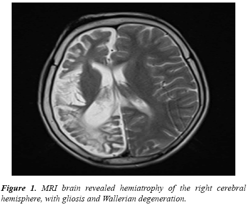
Hematological investigation revealed microcytic hypochromic anemia, renal function test, and liver function test was normal. MRI Brain was done, which revealed Hemiatrophy of the right cerebral hemisphere, with gliosis and Wallerian degeneration (Figure 1), characteristic of Dyke-Davidoff-Masson syndrome. EEG revealed generalized slowing and seizures activity and started on antiepileptic, valproate at 20 mg/Kg/day and eptoin was tapered and stopped because the child was having an intermittent seizure on eptoin which was already on maximum recommended maintenance dose. He responded well to drug therapy and physiotherapy and on follow-up showing improvement.
Discussion
In 1933 Dyke-Davidoff-Myke studied nine patients with peculiar clinical and radiographical findings like pneumatoencephalographic changes and seizure, facial asymmetry [2].
Dyke-Davidoff-Masson syndrome is characterized by hemiparesis, facial asymmetry, seizures, and mental retardation. CT and MRI features include cerebral hemiatrophy leading to dilation of sulci and cisterns and ex-vacuo dilation of the ventricle with compensatory contralateral calvarial thickening and hyperpneumatization of paranasal sinuses. The syndrome is of two types depending upon the etiology; Congenital or primary includes conditions like infection, coarctation of middle cerebral artery congenital anomalies or it could be due to lack of development of cerebral hemisphere on one side and idiopathic.
Secondary or acquired includes conditions like perinatal hypoxia, intracranial hemorrhage and infections, prolonged seizure, cranial trauma, cerebrovascular lesion [3,4]. In DDMS, calvarial changes occur only when brain damage is sustained before three years of age, but such changes may become visible as early as nine-months after brain damage. Brain growth failure compels inward redirection of nearby calvarial growth accounting for the compensatory calvarial changes like enlargement of the frontal sinus, increased width of the diploic space with elevations of the ipsilateral greater wing of the sphenoid, petrous ridge and planum sphenoidale [5,6].
Clinical presentation includes seizures, mental retardation, contralateral hemiparesis, facial asymmetry. Our patient presented with uncontrolled Diabetes Mellitus, with left-sided partial seizures, left-sided hemiparesis, and facial asymmetry. Birth history and antenatal history was normal. MRI brain showed hemiatrophy of the right-sided cerebral hemisphere with gliosis with Wallerian degeneration was seen.
This being a rare disease is likely to be missed and underdiagnosed. Characteristic clinical features, along with CT and MRI which display underlying gross pathologic changes as a result of prolonged atrophy has an important role in pointing towards the diagnosis. Knowledge of clinical features, risk factors, imaging features is therefore indispensable for making the diagnosis and for appropriate patient management.
References
- Sharma S, Goyal D, Negi A, Sood RG, Jhobta A, et al. Dyke-Davidoff Masson syndrome. Indian J Radiol Imaging. 2013; 16: 165.
- Abdul Rashid AM, Faeez Noh MS. Dyke-Davidoff Mason syndrome: A case report. BMC Neur. 2018; 18: 76.
- Uduma Felix U. Differential diagnoses of cerebral hemiatrophy in childhood: A review of literature with illustrative report of two cases. Glob J Health Sci 2013; 5: 195-207.
- Sharma S, Goyal D, Negi A, Sood RG, Jhobta A, et al. Dyke-Davidoff Masson Syndrome. Neuroradiology. 2006; 16: 165-166.
- Singh P, Saggar K, Ahluwalia A. Dyke-Davidoff-Masson syndrome: Classical imaging findings. J Pediatr Neurosci. 2010; 5: 124-125.
- Thakkar PA, Dave RH. Dyke–Davidoff–Masson syndrome: A rare cause of cerebral hemiatrophy in children. J Pediatr Neurosci. 2010; 11: 252-254.
