Current Pediatric Research
International Journal of Pediatrics
Down syndrome with PDA defect - A case report.
Kumar Sudhansh1*, Govind Singh Yadav1, Dundigala Narendar Kumar1, Motamarri Naga Sai Manikya Deepu1, Raj Wardhan1, Siddharth1, Saksham Srivastava1, Soni Kumari2
1Department of Pediatrics, T S Misra Medical College and Hospital, Lucknow, India
2Department of Prosthodontics, Patna Dental College and Hospital, Patna, India
- Corresponding Author:
- Kumar Sudhansh
Junior Resident, Assistant Professor Department of Pediatrics, T S Misra Medical College and Hospital, Lucknow, India
E-mail: sudhanshkumar9@gmail.com
Received: 24 September 2023, Manuscript No. AAJCP-23-114659; Editor assigned: 26 September, 2023, Pre QC No. AAJCP-23-114659 (PQ); Reviewed: 09 October, 2023, QC No. AAJCP-23-114659; Revised: 20 October, 2023, Manuscript No. AAJCP-23-114659 (R); Published: 27 October, 2023, DOI:10.35841/0971-9032.27.08.1185-1189.
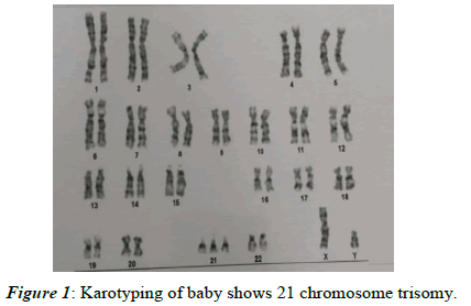
The Down Syndrome (47,XY,+21) is a rare chromosome abnormality. The aim is to describe the clinical features and diagnosis of rare event. A newborn boy was born to our unit at TS Misra Medical College & Hospital. He was born at 37 weeks' gestation via caesarean section due to Latent Labour. The boy was 49 cm in length, 2345 Gram in weight with head circumference of 31 cm. He had the characteristic features, including flat facial profile, flat nasal bridge, short thick neck, low-set ears, high palate, simian crease, epicanthal fold, slanted palpebral fissures, sandle gap. Hypotonia was noted as well. The Doppler echocardiography showed PDA defect (3 mm) with left to right shunt, and mild tricuspid regurgitation, with right atrium and right ventricle mildly enlarged. Cytogenetic analysis confirmed a karyotype of 47,XY,+21. Down syndrome is a rare disease. Karyotyping should be performed for all patients with suspected Down syndrome and in even babies born from young parents.
Keywords
Down syndrome human karyotype 47,XY,+21, Chromosomal abnormality, Congenital heart disease, Down syndrome, ECHO, Karyotyping.
Introduction
Down syndrome (Trisomy 21) is the most common chromosomal disorder named after Dr. John Langhan Down who first described it in 1866 [1]. Approximately 75% of conception with Trisomy 21 die inutero. Approximately 85% of infants survive upto 1 year [1].
The risk of having trisomy 21 increases with increasing maternal age [1]. Affected children have delayed physical growth, mental retardation, bone development and multiple congenital malformations.
Ten of the characteristic dysmorphic features are common in newborns with Down syndrome and are usually recognized soon after Birth-Hall’s criteria [2,3]. If 4 of the abnormalities are present 48% chance of being Down syndrome and if 6 abnormalities are present, the percentage of being Down syndrome raises to 89%.
• Flat facial features (90%)
• Slanted palpebral fissure (80%)
• Anomalous ears (60%)
• Hypotonia (80%)
• Poor moros reflex (85%)
• Simian crease (45%)
• Excessive skin at nape of the neck (80%)
• Clinodactyly (60%)
• Dysplasia of pelvis (70%)
• Hyperflexibility of joints (80%)
• Congenital heart disease is the most common cause of mortality [4,5]
• The incidence is about 40%-50% as compared to 0.4% in normal infants [6]. The following lesion are seen in Down syndrome
• Complete atrioventricular septal defect-37%
• Ventricular septal defect-31%
• Atrial septal defect-15%
• Partial atrioventricular septal defect-6%
• Tetralogy of Fallot-5%
• PDA-4%
• Others-2%
Case Presentation
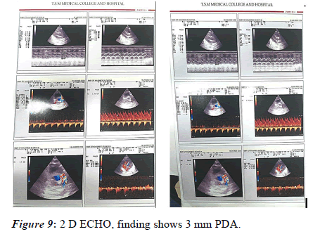
A newborn boy delivered to our Hospital and baby was kept in Neonatal Intensive Care Unit (NICU) in view of down phenotype and respiratory distress with Downes score 4. The boy was delivered to a gravida 2 and para 2 of 30 year-old mother and 35-year-old father. He was born at 37 weeks' gestation via caesarean section due to Latent labour. The Apgar score was 6,7 and 10 at 1, 5 and 10 minute respectively, and his birth weight was 2345 gram. Baby had signs of respiratory distress in form of tachypnea and sub-costal retractions with DOWNES Score 4, Humidified High Flow Nasal Cannula (HHHFNC) with flow 5 and FiO2 50 was started [6-8]. On auscultation baby was having Systolic Murmur for which 2D ECHO was done on day 2 of life the Doppler echocardiography showed PDA defect (3 mm) with left to right shunt, mild tricuspid regurgitation and right atrium and right ventricle mildly enlarged. For PDA defect IV Paracetamol administered for 3 days, Repeat 2D ECHO again done on Day 7 of life which showed tiny PDA defect (Figure 1).
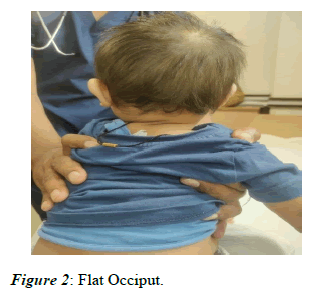
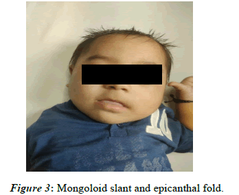
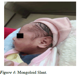
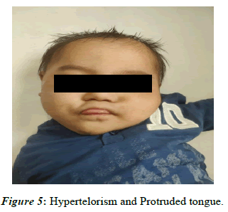
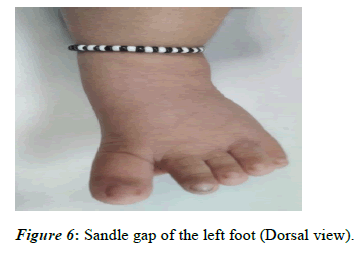
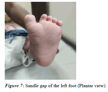
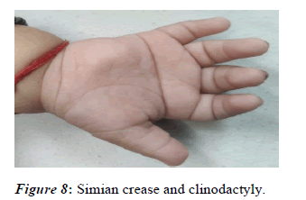
On Anthopometry examination, the boy was 49 cm in length, 2345 gram in weight with head circumference of 31 cm (<3 percentile). His respiratory rate was 60 times per minute without rales in the lung. No cyanosis was noted. He had the characteristic features, including flat facial profile, flat nasal bridge, short thick neck, low-set ears, high palate, simiancrease, sandle gap, clinodactyly, slanted palpebral fissures, hypertelorism, flat occiput and brachycephaly (Figures 2-8).
Generalised hypotonia was noted. Anterior fontanelle was 2×2 cm. The external genitalia were immature male with palpable testis in the scrotum. The dermal, ocular, thoracic examination were unremarkable. Chest X-ray showed normal findings in the lung (Figure 9).
Laboratory tests of liver, kidney and thyroid function were all normal. Cytogenetic analysis confirmed a karyotype of 47,XY, +21. Repeat 2D ECHO done in July 2023 showed closed PDA.
Discussion
The risk of having a child with trisomy 21 is highest in women who conceive after age 35 yr. Even though younger women have a lower risk, they represent half of all mothers with babies with Down syndrome because of their higher overall birth rate. All women should be offered screening for Down syndrome in their 2nd trimester by means of 4 maternal serum tests (free β- human chorionic gonadotropin (β-hCG), unconjugated estriol, inhibin, and α-fetoprotein). This is known as the quad screen; it can detect up to 80% of Down syndrome pregnancies vs 70%in the triple screen. Both tests have a 5% false-positive rate. There is a method of screening during the 1st trimester using fetal Nuchal Translucency (NT) thickness that can be done alone or in conjunction with maternal serum β-hCG and Pregnancy- Associated Plasma Protein-A (PAPP-A). In the 1st trimester, NT alone can detect ≤ 70% of Down syndrome pregnancies, but with β-hCG and PAPP-A, the detection rate increases to 87%. If both 1st and 2nd-trimester screens are combined using NT and biochemical profiles (integrated screen), the detection rate increases to 95%. If only 1st-trimester quad screening is done, then maternal serum α-fetoprotein (which is decreased in affected pregnancies) is recommended as a 2nd-trimester followup [1].
Clinical features of Downs syndrome include, flat nasal bridge upward slanting palpebral fissures, brushfield spots, small ears, small nose, excessive skin at the nape of neck, common medical problems include hearing problems, vision problems, cataracts, obstructive sleep apnea, otitis media, congenital heart disease, thyroid diseases, seizures, hematologic problems [9].
In the present case Clinical features of the patient were similar to the previous studies such as Epicanthal folds of eyes, depressed nasal bridge, protruded tongue, short neck, hypertelorism, mongoloid slant, Sandle gap, Simian crease, Clinodactyly, Flat occiput, Brachycephaly.
In this present case no NT (Nuchal Translucency), NB (Nasal Bone) scan was not done neither Level 2 USG was done. Diagnosis of Downs syndrome primarily includes screening test which are non-invasive test such as ultrasonography. Definitive diagnostic test includes Karyotyping, Cultured fetal cells such as Chorionic Villus Sampling (CVS), Aminocentesis [10]. During the 2nd and 3rd trimisters of pregnancy Percutaneous Umbilical Blood Sampling (PUBS) is a method for obtaining fetal blood along with ultrasonography [11].
Conclusion
One of the hallmarks of Down syndrome is the variability in the way that the condition affects people with DS. With the third 21st chromosome existing in every cell, it is not surprising to find that every system in the body is affected in some way. However, not every child with Down syndrome has the same problems or associated conditions. Parents of children with Down syndrome should be aware of these possible conditions so they can be diagnosed and early and timely intervention. Timely surgical treatment of cardiac defects during first 6 months of life may prevent from serious complications. Feeding problems and failure to thrive usually improve after cardiac surgery. To improve our understanding of the disease numerous theories have been put forward. This case report highlights different aspects of this condition which might help in the diagnosis of the condition. PDA defect is one of the least common congenital cardiac defects in patients of Down Syndrome. The presence of cardiac defect such as PDA can be treated medically if not resolved surgical correction can be done. A balance diet and regular exercise are needed to maintain appropriate weight. Most males with Down syndrome are sterile, but some females have been able to reproduce, with a 50% chance of having trisomy 21 pregnancies. Two genes (DYRK1A, DSCR1) in the putative critical region of chromosome 21 may be targets for therapy. In this case, for PDA defect IV Paracetamol was administrated for 3 days, repeat 2D Echo before discharge of baby from NICU showed only tiny PDA defect. Repeat 2D ECHO in July 2023 showed PDA has closed. A Down syndrome child should have regular check-ups.
References
- Kliegman RM. Geme J St. Nelson Textbook of Pediatrics. 21st Edition 2019.
- Lott IT, Dierssen M. Cognitive deficits and associated neurological complications in individuals with Down's syndrome. Lancet Neurol 2010;9(6):623-633.
- Kent L, Evans J, Paul M, et al. Comorbidity of autistic spectrum disorders in children with Down syndrome. Dev Med Child Neurol. 1999;41(3):153-158.
- Huang XY, Tang LF, Zou CC, et al. Chromosome analysis in 8158 pediatric patients: An experiment of 29 years from Hangzhou, China. Endocrinologist 2010;20(4):179-181.
- Xu F, Zou CC, Liang L, et al. Case Report Ring Chromosome 15 Syndrome: Case Report and Literature Review. HK J Paediatr 2011;16(3):175-179.
- Caputo AR, Wagner RS, Reynolds DR, et al. Down syndrome: Clinical review of ocular features. Clin Pediatr. 1989;28(8):355-358.
- Kazemi M, Salehi M, Kheirollahi M. Down syndrome: Current status, challenges and future perspectives. Int J Mol Cell Med 2016;5(3):125.
- Tarique M, Afreen S, Jalal M. Down's syndrome: A case report. Inter J scie res 2008: 638-634.
- Natoli JL, Ackerman DL, McDermott S, et al. Prenatal diagnosis of Down syndrome: a systematic review of termination rates (1995–2011). Prenat Diagn. 2012;32(2):142-153.
- Walker BR, Colledge NR. Davidson's principles and practice of medicine e-book. Elsevier Health Sci 2013.
- Dierssen M, Ortiz-Abalia J, Arque G, et al. Pitfalls and hopes in Down syndrome therapeutic approaches: In the search for evidence-based treatments Behav Genet. 2006;36:454-468.
